Weber Fracture Treatment, Surgery - Weber Fracture Types Healing Time, Recovery
As indicated by an investigation on bone crack, the lower leg is one of the every now and again harmed joints in the body. Patients with this type of fracture have to undergo lateral x-ray and AP for fracture diagnosis in order for the surgeon to know how to manage the injury. There are different types of ankle fractures that have to be analyzed to know how to heal each type. Proper classification of the fracture will also help the doctor analyze the stability of the fracture injury and the extent of the damage. For better understanding, the Weber classification of ankle fractures will be combined with the Lauge-Hansen system. The Weber A, B, C fracture fracture evaluation would be done easily then.In the case of other types of fracture such as Salter Harris fracture, there are different types of fractures that happen on the growth plate of kids. This Salter Harris fracture classification also uses another system. On the other hand, Schatzker tibial plateau fracture is under another classification system. Schatzker fracture classification is a system used to categorize tibial fractures.

The Weber Classification System
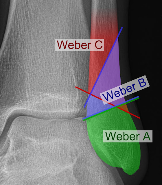
This is based on the syndesmosis of the injury or the degree of articulation of the fracture of the ankle. The Weber fracture classification system has become famous due to its simplicity. This is how it classifies ankle fracture.- Weber A fracture. The fracture happens below the syndesmosis and the broken bone is intact.
- Weber B fracture. The fracture is considered transsyndesmotic with a partial break of the syndesmosis.
- Weber C fracture. The fracture happens just above the syndesmosis which is totally ruptured. This is also characterized by the instability of the fractured ankle.
This is based on the trauma mechanism of the fractures. The types of ankle fracture are determined after radiographs are obtained from the fracture. The severity of the fracture will be analyzed. Thus, the Lauge-Hansen system is already a big help to the doctor in analyzing the instability and damage of the ligamentous injury.
The criteria of this system’s classification of ankle fractures are:
- Direction of the force relative to the foot, which could be adduction (20%) and exorotation (80%).
- Position of the foot during the occurrence of the injury, which could be pronation (20%) or supination (80%)
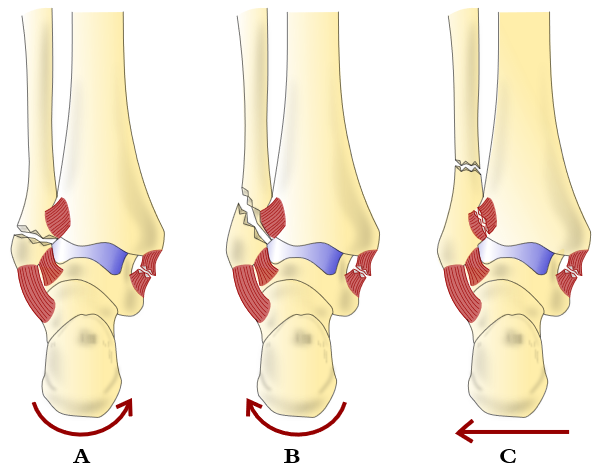
Weber A Ankle Fracture – Lauge Hansen SA
This is the simplest type of Weber ankle fractures. The diagnosis of this Weber fracture and its treatment can be done simply. This type of Weber bone fracture accounts for 20% to 25% of all the ankle fractures. The injury could have happened when the foot is in supination and the talus is hit by an adduction force. The fracture of ankle will likely be felt first on the lateral side with a lot of tension. Here is how the Weber A fibula fracture or similar type A fracture has happened:- The supination on the foot causes a tear on the lateral collateral ligament. This could also result to an avulsion fracture on the part just below the tibial plafond and the syndesmosis.
- The medial malleolus is pushed off vertically when the foot moves on a talar tilt. This makes the Weber A fracture ankle unstable.
It should be noted though that that malleolar ankle fracture are comparable to a ligamentous injury. Another fracture on the tibia, particularly an avulsion fracture, would be the tillaux fracture. This happens when the anterior syndesmosis happens to get attached. However, this type of fracture rarely happens.
Weber B Ankle Fracture
This accounts for 60% to 70% of all the Weber fractures, making it the most common among the types of fracture of ankle. In this fracture, the foot is still in supination while the talus got hit by an exorotation force, which happens when the lower leg does an endorotation. This is how Weber type B fracture happens stage by stage:- The first break on the bone during a Weber B fibular fracture or similar fracture would be on the lateral side of the ankle. This is due to the maximum tension that has dominated this side. The tidiofibular ligament breaks first when the talus does an exorotation.
- With the foot in supination, the firm hold of the lateral collateral ligament would hold the lateral malleolus in place. As a result, it would break with just a simple move. Thus, when the talus rotates, the Weber B fibula fracture becomes worse and cause a seemingly spiral break on the bone. This is also triggered when the lateral malleolus moves from anterior to posterior after being pushed off. The weber B distal fibula fracture usually happens above the ankle, just a few centimeters from it, and continues to break proximally.
- After the displacement of the lateral malleolus after being pushed off, the posterior tidiofibular ligament would break. This is also considered as the avulsion fracture of the malleolus tertius.
- As the talus moves to the posterior, the medial side of the ankle would be under more pressure. This will result to a rupture of the detoid ligament.
Generally, the supination of this type of Weber fractures usually occurs in a clockwise manner. The exorotation injury of the Weber fibula fracture happens in the following manner:
- Tearing of the anterior tibiofibular ligament
- Oblique breaking of the distal fibula
- Tearing of the posterior tidiofibular ligament
- Tearing of the medial collateral ligament
The diagnosis of this type of Weber injury might be a bit complicated. The result of the x-ray might not show any fractured ankle because the broken bones and ligaments would align back together. Thus, further diagnostic tests might be performed to find the Weber ankle fracture especially if the pain persists. This is one of the ever present fracture symptoms that should tell the doctor and the patient that something is definitely wrong after an injury. The diagnosis should be done correctly in order to know how to treat Weber fracture effectively. An effective Weber fracture treatment would definitely be a big help on reducing the length of the fracture healing time.
Weber C Ankle Fracture
This accounts for 20% of the fractures that occur on the ankle. Weber C fibula fracture happens when the foot is planted on the ground in pronation while the talus is hit with an exorotation force. This Weber C fibular fracture happens in the following stages:- The injury will first happen on the ankle’s medial side. The tension is at the strongest in this area which immediately causes it to break. This will result to the tearing of the medial collateral ligament or to the avulsion fracture of the medial malleolus.
- Upon being freed from its medial ligament, the talus will rotate externally. However, as the foot is in pronation, the fibula would move away from the tibia with the absence of tension on the lateral side. This will result to the tearing of the anterior syndesmotic ligament.
- A distal movement will happen to the fibula while it remains fixed in is position. The interosseus membrane will tear at this point and the fibular shaft will break above the syndesmosis. However, x-ray tests might not show the fibular fracture.
- The posterior syndesmotic ligament would tear apart.
The Weber C fracture ankle also happens in a clockwise manner, just like type C. However, this fracture involves an pronation exorotation injury on the fractured ankle bone.
- Tearing of the medial collateral band
- Tearing of the anterior tidiofibular ligament
- Fractures of the fibula in a transverse manner
- Tearing of the posterior tidiofibular ligament
An experienced doctor would know how to interpret each of these types of injury to determine how to treat Weber fracture quick. The surgeon might determine if fracture surgery is necessary or not. In some cases of broken ankle bones, the patient would be asked to wear a fracture cast for a few weeks. This way, the recovery time would be determined and the patient would then be able to make plans around this period. To achieve complete recovery, rehabilitation should be done. A properly done rehab can be facilitated by a physical therapist, who can help you achieve complete recovery at the right Weber fracture recovery time.

I want to thank Dr. Uduehi for helping me shrink/cure multiple FIBROID. It’s been a year since I got free from the problem but I decided to wait to see if they will grow back and luckily no sign of it at all. I normally experience heavy menstrual blood, Pelvic pressure or pain, Backache, Frequent urination ETC. And now, all the symptom or pains are no more. I went to the hospital again to check and was diagnosed fibroid free. All thanks to doctor Uduehi. You too can be free, contact him now for a cure through: +234-708-487-8384 (uduehiherbalcare@gmail.com)
ReplyDelete