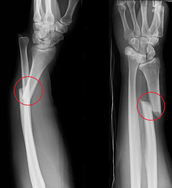
A bone fracture is a condition of the bone that has a crack or break. The bones in the human body need calcium phosphate in order to grow dense and strong. As a person reaches the age of 30, the bone density reaches its highest level. It is at this age that the bones are at their strongest. However, they are no match to a very strong external force that might cause a crack or break on the bones. There are complete and incomplete fractures, based on how the bones are broken or cracked. When it happens that the fractured arm, wrist, forearm, or any bone is still intact, the result would be an evident bend in the bone.
There are different types of bone fractures. Some would occur on the hips, wrist, arm, foot, neck, spine, skull, and many other bones in the body. One such type one of fracture of arm is the Galeazzi fracture. In 1929, the Frenchmen dubbed this fracture as the anti-fracture of the glenoid. This was originally discovered by Cooper, a doctor in the 19th century, but the credit goes to Dr. Galeazzi who studied this fracture further in the 1930s. Galeazzi made a detailed description of the fracture and came to the idea that thumb strength traction would be one of the ways on how to heal this injury. This fracture refers to a break in the radius bone, which can be located in the lower part of the arm just on the area close to thethumb. A dislocated distal radial ulnar joint also known as DRUJ is often associated with the Galeazzi bone fracture.
Galeazzi fractures are less common compared to other types of fracture injury on the arm. According to studies, this fracture accounts for three to seven percent among all the arm fractures. It was also observed that men are more prone to suffer from Galeazzi fractures than women. There are closed and open fractures, with the latter being more dangerous than the other due to their higher risk to get infected. A person with an open Galeazzi injury should obtain the quickest treatment possible. Galeazzi fracture treatment is urgent in the case of any open fracture, with the exception may be of the presence of some even more severe injuries.
History
In 1842, the world first heard of the Galeazzi fracture as it was defined by Cooper. However, a more detailed work on this arm fracture was done by the Italian Ricardo Galeazzi (who was born in 1866 and died in1952) while he was employed at the Instituto de Rachitici in Milan as a surgeon. His work on the broken arm bones came from his specialization on congenital hip dislocation. This particular study reported about 18 fractures with different patterns. These fractures were then named after him in his honor.
In 1941, the Galeazzi’s fracture was also called the fracture of necessity, because it demands immediate medical attention and treatment right after its occurrence. Fracture surgery is most likely the solution that is recommended by doctors who would examine patients with Galeazzi fracture wrist.
Pathophysiology
When fracture diagnosis, with the use of X-ray, has determined that Galeazzi injury does exist, the patient should be warned about some other forces that would cause deformity of the injured bone. A deformed and fractured arm bone would be an even bigger problem than any other forms of fractures. Thus, Galeazzi fracture in children should be carefully monitored by parents. Children tend to be naturally restless due to the high level of energy that hey have. You will rarely find children who will just stay seated in one corner when you tell them too. In a few seconds after they have behaved, they would find other ways to have fun and a trip or a heavy fall would likely change the scene to an accident. The forces that can cause deformed Galeazzi-injured bones are (1) pronator quadriceps, (2) brachioradialis, (3) thumb extensors, and (4) natural weight of the hand. Unlike other forces and movements in most cases of fractures, immobilization using a brace would be effective but this does not seem to work in a Galeazzi injury.
Fracture Symptoms
A case of Galeazzi fracture dislocation can be identified with the help of certain symptoms. Pain is an ever present symptom in almost all types of fractures. This is why medicines for pain relief are needed to help remove the feeling of discomfort from the patients. Swelling is bound to happen as well. The forearm fracture can be confirmed using radiographic evaluation. There are also bound to be some forearm trauma that will be felt by the patient.
Treatment
On the issue of how to heal Galeazzi fracture quick, there are different treatment options that are available. This type of fracture can be open or closed but the former would need immediate surgery to have stability for the DRUJ. These fractures might also come with some cracks or breaks on the other bones in the forearm. The surgery is still considered as the best treatment option to these cases.
A surgical procedure to treat Galeazzi fracture in adults would consist of reduction. This is the process of stabilizing the broken bones with the help of surgical pins. Stability is necessary for fracture healing time to start. Without this factor, recovery time for a Galeazzi injury would all the more be prolonged. The pins are to be placed in the fractured bones in the arm for about four weeks or a month. However, even after the pins are removed, a cast cannot be avoided with the thought that immobility has been achieved with the use of pins already. This is because a restrictive fracture cast or splint has to be placed on the fractured bone for another four weeks, in most cases. This is why it would take a long Galeazzi fracture healing time for complete recovery.
However, treatment of Galeazzi fracture or fractures of the arm in little children is likely to avoid the use of surgery. More doctors would favor treating the injury in closed form. Adults need open surgery in order to avoid further fracture complications and restrictions in the future. Without immediate surgical treatment, the DRUJ is likely to sustain more damage and it might be harder to move the injured bone even after the supposed healing period. In the case of the injury in children, their bones are still going to develop. Thus, they tend to heal faster and in a more predictable manner than those of the adults. However, severe cases might prompt the doctor to recommend surgery for immediate treatment.
After the surgery, recovery would likely happen within two months. Rehabilitation is necessary in some cases, especially with severe fractures. This period is likely to start after the restrictive braces, cast or splint is removed from the injured area. There are patients who would also be asked to take prescribed emedicine if some discomfort can still be felt. During rehab, some stretching and strengthening exercises have to be performed, but all the routines should go through the evaluation of the doctor first. There are still some movements that should not be performed by the newly healed bone. A physical therapist would be a big help in building up strength in the bone that has been immobilized for about two weeks. The exercise routines will help the healing bone learn to move again after it was placed in braces.
As per observation in a certain study, the healing process and the time it takes for the process to get completed depends on several factors. These factors would include the age of the patient, extent of the damage of the fracture, and how the surgery was performed. Thus, it is essential that a good surgeon should be found. About 40 percent of the attempts to heal the injured bone would encounter some complications. However, these risks and complications are even more probable when surgery is not done in the case of adults who have Galeazzi injury. In fact, surgery might also be required among those who suffer from glenoid fracture. Other patients who have sacral fracture in severe from have to brave a surgery too. Insufficiency fracture, a type of stress fracture, also has surgery as an option. This goes the same for those who have intertrochanteric fracture as well.

Comments
Post a Comment
Please do not enter any spam link in the comment box.