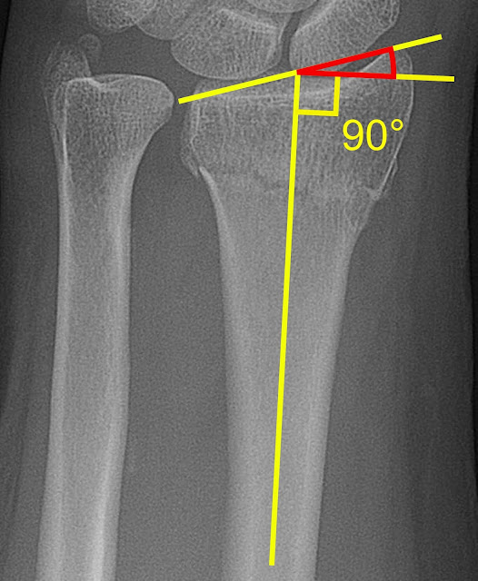
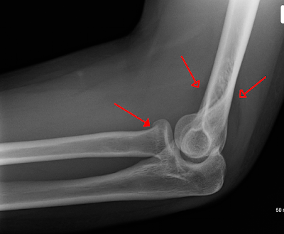
The two long bones of the forearm is the radius and the ulna and they are both very important for proper motion of the wrist and the elbow. The radius is larger than the ulna and it is positioned on the front side of the arm that is on the side of the thumb. However, although the radius is a strong and large bone, it is also prone to fractures. Radial fractures usually occur when someone falls hard with an outstretched arm. The tendency of the person to cover his or her body to break the fall can cause the radius to break because the forearm will be absorbing all the tension and weight of the person during a fall.
Types of Radial fractures
Some of the most recognized radial fractures are:
• Proximal radial fracture. This is a fracture that is near the radial head or radial neck. It can occur due to direct or indirect injury to the forearm or elbow joint. Because the elbow is a hinge joint that joined the ulna and the radius, when there is a sudden impact to the elbow, the strong energy will fracture the elbow and then the excess energy will travel to the nearest bone and fracture the radius.
• Distal radius fractures. This is caused by a fall with an outstretched hand or can be the result of axial forces or direct impact. Some of the associated anomalies within this fracture can be any of the following:
• Barton’s volar fracture
• Barton’s dorsal fracture
• Colles fracture
• Chauffeur’s fracture
• Intra-articular fractures
• Radial Shaft fractures: This is an isolated case of radial fracture and it is usually associated with fracture to the ulna.
Some other common types are radial head fracture, styloid, neck and wrist.
Radial Head Fracture Diagnosis
If there is the deformity of the radius, the physician will conduct immediate clinical diagnosis on the injury and will confirm his analysis with the result of the X-rays. He will also conduct a differential diagnosis that will include the scaphoid and the wrist because fractures of the radius are always accompanied by wrist fractures. If the fractures are not clearly seen on X-rays, delayed X-rays, MRI or CT scan will confirm the diagnosis further. Rehabilitation or rehab is necessary.
Radial Fracture Causes
Although radial fractures are not unusual, falls with an outstretched hand, vehicular accident, skiing accident, bike accidents can cause radial fractures. The most affected bone however is the radius and not the ulna because the radius is the bigger bone that usually absorbs most of the shock during falls. However, when the ulna is also fractured, this case is called the distal ulna fracture and it is quite hard to treat than simple radial shaft fracture.
Radial Fracture Symptoms
Radial fractures square measure easy to recognize as a result of as before long because the injury happens, symptoms can seem.
Here are the common symptoms:
• There would be unbearable pain on the injured area and the pain can travel to the upper arm.
• Bruises will show up especially around the broken area
• Swelling will also be there hours after the injury
• There will be tenderness on skin
• If the arm is moved there will be unbearable pain within
• There would be deformity for serious fracture
Radial Fracture Treatment
Radial fractures can be under two categories, namely, open and closed fractures. For open radial bone fractures, the broken bone protrudes through the skin. This is a serious case of bone fracture and requires immediate medical attention because anytime the infection can set in. The person could also go into shock due to blood loss. In the hospital, surgery is needed to clean and disinfect the wound and reconnect the damaged tissues and tendons. Bones will be reattached with the use of metal wires and plates if several fractures occur. The healing can take longer to complete.
In incomplete fractured radial, the fracture has kept the bone in place and no bone fragments have been caused. In this case, the doctor may put the arm in cast plaster or in splint to immobilize it until the bone reconnects and heals itself. Pain relievers are always part of the treatment because from time to time, the fractured bone will ache especially when the weather is cold. Rehabilitation always follow right after the healing of the bones. Physiotherapy can help a lot.
Radial Fracture Healing Time & Recovery Time: It requires more than 8 weeks.
Prevention of Radial Bone Injury
A good nutrition to prevent radia bone injuries is one of the best advices for bone fracture prevention. Calcium and Vitamin D rich foods are the best source of bone strengthening minerals. You can also minimize bone fracture especially fracture on the forearm if you will wear pads on your arm when doing activities that could put your arms at risk. However, better avoid activities or sports that are too much for your bone structure. If you have a light and small frame, you will always be prone to bone fracture. Strengthen your muscles and bones also with exercises and always do warm-ups before engaging in sports.
When to Call A Doctor
Radial fractures are common because it is our instinct to use our arms in protecting ourselves during falls or to stop blows from coming to our bodies. Therefore, if you see someone suffered from a fractured forearm, call the emergency hotline as soon as possible. For your information, radial nerves are crucial nerves that supply blood to the arm. If the nerves could be damaged, there will be more complications to the forearm.


Comments
Post a Comment
Please do not enter any spam link in the comment box.