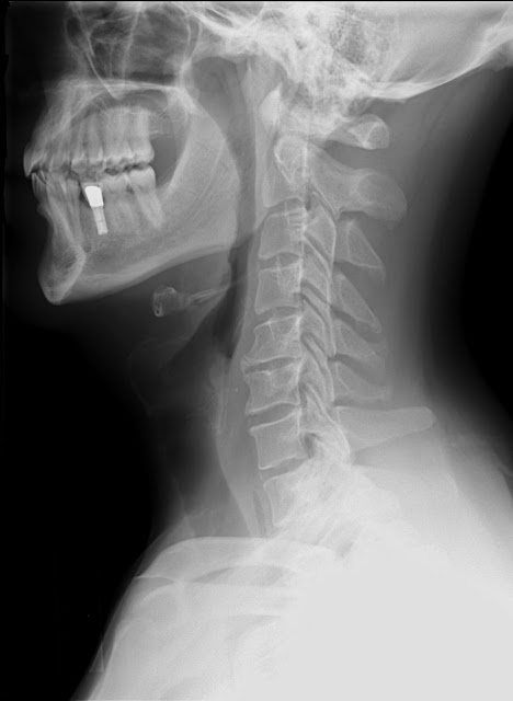
Unstable fractures are actually types of fractures which, despite being treated with bone reduction method still slipped out of their proper places. An unstable fracture is usually the result of an incident wherein a high level trauma had an impact to the bone, making the bone hard to realign with the use of simple treatments. Here, the main fragments of the fragmented bone are too many so that it usually results to a nonunion or a very slow union of bones.
Unstable Fracture Types
Unstable fractures are those fractures, which slip even after reduction is done. The most common types of unstable fractures are the following:
• Unstable Pelvic fractures: This is caused by high-energy trauma like the ones people get from vehicle accidents. The pelvic is a dangerous area when afflicted with unstable fractures because this type of injury can cause hemorrhage and multiple failures of important organs found near the pelvic site. Most pelvic unstable fracture cases are treated using multidisciplinary approach.
• Flexion teardrop fractures of the spine: This unstable fracture of the spine is brought about by too much damage on the bone and its ligaments. If this injury is kept on its unstable stage, the individual can experience a decrease in height due to the affected vertebral body.
• Unstable elbow fractures: This is considered as one of the most severe cases of bone injuries. Most cases of this injury require surgery because they are one of the most challenging fractures to correct. During the surgery, the surgeon is faced with the challenge of properly scrutinizing the dislocation of the elbow so as to avoid missing other associated injuries the fracture has caused.
There can be unstable ankle, unstable knee, joint, neck, back, spine, hip, unstable shoulder and pelvis.
Diagnosis
Diagnosing unstable bone fracture starts with a brief interview and the conduct of physical examination to the patient to identify whether the fractured bone is really unstable or not. The physician will ask the patient about the history of the injury. Physical examination will be conducted followed by X-rays. If there is the possible occurrence of more damage to the bone and its surrounding tissues, an MRI or CT scans will be recommended so that a full-scale imaging will be shown. Rehabilitation or rehab is very important.
Unstable Fracture Causes
• High-energy trauma from vehicle accidents
• Falling from heights
• Slipping and falling on the elbow (for elbow fractures)
• Not having enough rest time after reduction
Unstable Fracture Symptoms
• Extreme pain
• Swelling
• Bone sticking on the skin
• Numbness
• Having a sore feeling on the area
Unstable Fracture Treatment
Just like in diagnosis, the type of treatment for unstable fractures is dependent on the body part where the injury is located. Before, unstable pelvic ring injuries were only treated with the use of conservative treatments together with a lengthy bed rest. Nowadays, using the operative or internal fixation method is more preferred because unstable pelvic fractures have high possibilities of damaging the organs near it.
Through the use of surgery, internal bleeding or ripped tissues that cannot be sensed by bone scans can be given attention. Moreover, some fractures are aided with the use of external fixation tools like slings and casts. For unstable spinal fractures, the use of a metal abdominal brace might be needed to immobilize the spine.
Surgery requires the use of tools like pins, K-wires, and plates to align the unstable bones on their right places. For most unstable fractures, this method is more advantageous because the probability of developing another unstable injury is lesser once the fracture is already administered with internal fixation.
Unstable Fracture Healing Time and Recovery Time: It requires more than 8 weeks.
Prevention of Unstable Bone Injury
To prevent a fracture or unstable bone injury from turning into an unstable injury, having enough rest after the reduction was applied is the most effective thing. A patient who had just undergone a reduction treatment should keep his injured area as immobile as possible and away from extreme temperatures. If the doctor didn’t prescribe physical therapy, refrain massaging or administering any type of exercise on the injured part because it can cause the slipping/misaligning of the bones.
When to Call a Doctor
Experiencing extreme pain after a couple of weeks since having reduction treatment is not normal so a situation like this calls for immediate medical attention. Look for symptoms of possible bone injuries and if you see any such as swelling, bleeding, and bruising, call medical help right away. The bad thing about unstable fractures is that sometimes they are more painful than new fractures and are usually hard to treat especially if there is infection in it. Ask your doctor on how to prevent unstable fractures because some patients are being careless, which worsens their injury even more.

Comments
Post a Comment
Please do not enter any spam link in the comment box.